What is an Ovarian Dermoid and Why Does It Grow Teeth!?

My first experience with an ovarian dermoid tumor was in medical school. I saw a picture in our text book and was immediately repulsed – how can something that creepy be a real-life finding?!
Though the tumors continue to be one of the stranger pathologies I deal with, the repulsion-factor has subsided. So, what in the heck is an ovarian dermoid and why would an ovary have teeth inside it?
What I’m calling a “dermoid” is just another name for the most common type of ovarian germ cell tumor, in textbooks it will usually be called a mature teratoma. Dermoid came about in modern medicine after the discovery that dermal elements frequently predominate the tumors.
The origination of the word teratoma is from the Greek word teras, which means monster. Since they can contain fully formed teeth, bone, hair, and other weirdness, it is not hard to figure out why this would have been the name people gave it way back before we knew about how the out-of-place tissue got there.

If you haven’t read the book “Brain on Fire” by Susannah Cahalan you need to get your hands on it. We read it for book club in residency and all the spouses enjoyed it as well, it’s definitely not targeted at medical audiences. Dermoid tumors are occasionally (rarely) associated with an unusual condition called NMDA-Receptor Encephalitis and this is a fascinating and well-written memoir by a New York Post reporter documenting her long and life-threatening journey to this diagnosis.
Pathophysiology, Diagnosis, Treatment…
Dermoids are neoplasms which arise from primordial germ cells and there are several types of teratomas. For board exam purposes, here’s some broad, quick, key-word references:
-
Mature Teratoma = Common = Benign
– AKA: Dermoid/Cystic Teratoma -
Immature Teratoma = Rare = Malignant
– AKA: Teratoblastoma or Embryonal Teratoma
Teratomas in general are defined by the fact that they arise from a single germ cell, meaning they have the capability to differentiate into any of the germ cell layers and frequently contain all three (endoderm, ectoderm, mesoderm). This is why they’re so weird – they are comprised of tissue that would not normally be found in the ovary!
There’s also a less common type of teratoma called “monodermal teratoma.” This tumor differentiates to one special tissue, most commonly thyroid tissue, which is called struma ovarii and can present as thyroid storm!
What’s the most common tissue in an ovarian teratoma?
- Ectodermal Elements
– Skin
– Sebaceous & Sweat Glands
– Hair
– Teeth
How in the world does this happen?
- A single cell in an oocyte gets a wild hair and goes rogue, this is the most common theory
– So, genetically the DNA is 46, XX
– Embryonic tissue can develop, but they do not undergo complete embryogenesis
How do they present?

- Ovarian Torsion
– Patient presenting to the Emergency Dept with sudden onset abdominal pain and an adnexal mass is the most common way we find these. About 15% of Dermoid Cysts will eventually cause ovarian torsion. This happens because the fatty elements, which are low density, tend to make up a large portion of the cyst and this allows the mass to “float” in the abdominal cavity. Thus, they can easily roll/twist along the Infundibulopelvic (or Suspensory) Ligament of the ovary.
So, what if they are malignant?
- I know, I just said they aren’t – and about 99% of the time they’re not, but they can be. If a mature cystic teratoma IS malignant, it will usually be a squamous cell carcinoma.
– Why? Refer back to the most common tissue type – skin! - Skin cancer in the ovary…yup.
How are they treated?
- If symptomatic, treatment is via surgical removal with ovarian cystectomy or oophorectomy. Asymptomatic ovarian dermoids can be removed to prevent torsion or left in hopes of avoiding surgery.
What do they look like?

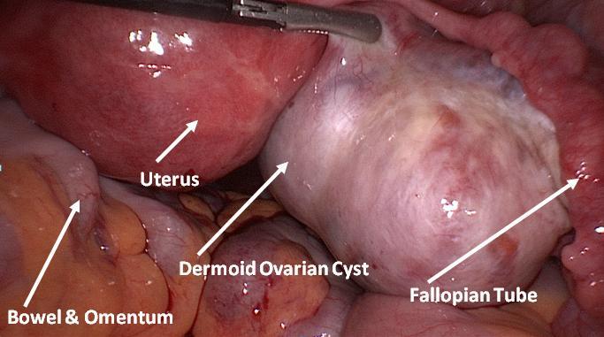
- Here’s a great picture from Queensway Gynecology. Click on their link to see really cool videos of laparoscopic surgical removal of a dermoid cyst or click here or here or here to see some of the more interesting teras-esque internal pathology shots.
